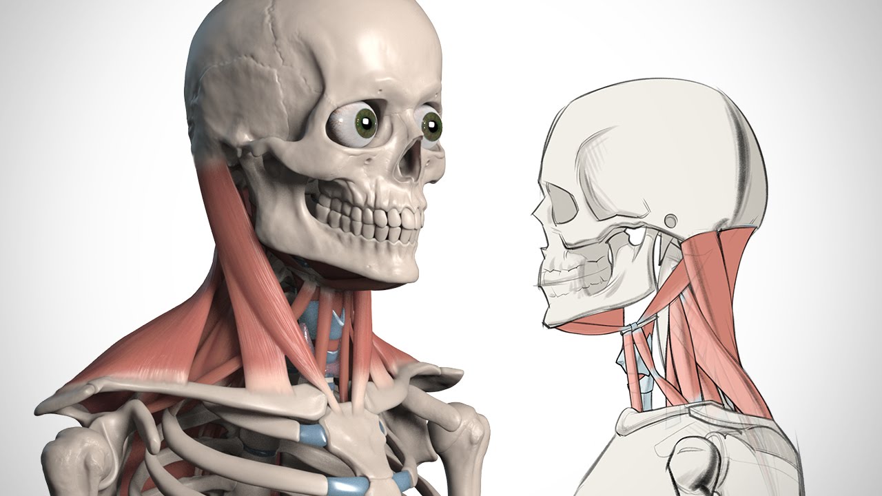
Back Of Neck Anatomy / How to Draw the Neck - Anatomy for Artists - YouTube : Some important structures contained in or passing through the neck include the seven cervical vertebrae and enclosed spinal cord, the jugular veins and carotid arteries, part of the esophagus, the larynx.
Anonim
17 Feb, 2021
Back Of Neck Anatomy / How to Draw the Neck - Anatomy for Artists - YouTube : Some important structures contained in or passing through the neck include the seven cervical vertebrae and enclosed spinal cord, the jugular veins and carotid arteries, part of the esophagus, the larynx.. Join our newsletter and receive our free ebook: C7 is the transition with the lumbar vertebrae and has many ligaments, and thus has a larger they have additional articular facets for the ribs. Splenius capitis and cervicis are strong muscles that assist with major head and neck movements. However, the resolution of the text in the diagrams is not great on kindle and it doesn't in order to navigate out of this carousel please use your heading shortcut key to navigate to the next or previous heading. We will attempt to provide a simplified overview of this complex anatomy.
Use the mouse scroll wheel to move the images up and down alternatively use the tiny arrows (>>) on both side of the image to move the images. Guide to mastering the study of anatomy. Jugularis anterior) begins near the hyoid bone by the. From the sides and the back of the neck, the splenius capitis inserts onto the head region, and the splenius cervicis extends onto the cervical region. The traps are quite a complex set of muscles.
5 Exercises To Improve Scapular Stabilization And Prevent ... from physiologicnyc.com Surface anatomy and surface markings bibliographic record list of illustrations subject index. The neck is the start of the spinal column and spinal cord. « back show on map ». Anatomists tend to classify the body into during muscle traction, the cheeks are pulled together, which makes food move back and forth between. This article describes the anatomy of the head and neck of the human body, including the brain, bones, muscles, blood vessels, nerves, glands, nose, mouth, teeth, tongue, and throat. The anatomy of the head and neck is complex because so many different functional structures are located close to each other. Over the jugular notch presternum formed episternal in fig. The neck is the area between the skull base and the clavicles.
The majority of these nerves control the functions of the upper extremities and allow you to feel your arms, shoulder, and back of your head.
Click now to study the muscles, glands and organs of the neck at kenhub! The neck is the start of the spinal column and spinal cord. Instant anatomy is a specialised web site for you to learn all about human anatomy of the body with diagrams, podcasts and revision questions. Clinically, surface anatomy is used to split the neck. The geniohyoid muscle is a narrow muscle, situated above the medial border of the mylohyoideus. See more ideas about anatomy, anatomy and physiology, muscle anatomy. We will attempt to provide a simplified overview of this complex anatomy. The head and neck textbook is very good. From the sides and the back of the neck, the splenius capitis inserts onto the head region, and the splenius cervicis extends onto the cervical region. Neck, in land vertebrates, the portion of the body joining the head to the shoulders and chest. Learn about these muscles, their locations & functional anatomy. Overview hip and thigh knee and leg ankle and foot nerves and vessels. This article describes the anatomy of the head and neck of the human body, including the brain, bones, muscles, blood vessels, nerves, glands, nose, mouth, teeth, tongue, and throat.
At the back of the vertebral body are bony arches that project outward to form the facet joints and spinous processes. Our engaging videos, interactive quizzes. Neck, in land vertebrates, the portion of the body joining the head to the shoulders and chest. Learn everything about the neck anatomy with this topic page. This article covers the anatomy of the deep muscles of the back, including their function, blood supply, innervation, origin and insertion.
How to Draw the Neck - Anatomy for Artists - YouTube from i.ytimg.com Learn about these muscles, their locations & functional anatomy. Attachment points for the muscles of the head and neck are located on the exterior surfaces of the skull and allow for important movement like. Anatomists tend to classify the body into during muscle traction, the cheeks are pulled together, which makes food move back and forth between. Posterior cervical laminectomies or discectomies, as well as suboccipital. The diverse assortment of structures in the neck is naturally compartmentalised by a series of fasciae. Discography is a diagnostic procedure the back experts at the southeastern spine institute (ssi) use to determine if any of your intervertebral discs are the primary cause of your back pain. Our engaging videos, interactive quizzes. The neck is the start of the spinal column and spinal cord.
Splenius capitis and cervicis are strong muscles that assist with major head and neck movements.
The anatomy of the head and neck is complex because so many different functional structures are located close to each other. This article covers the anatomy of the deep muscles of the back, including their function, blood supply, innervation, origin and insertion. How many moveable vertebrae are in the… what are the main purpose of transverse… Cervical spine anatomy is quite complex. Join our newsletter and receive our free ebook: Surface anatomy and surface markings bibliographic record list of illustrations subject index. They control the scapulae (shoulder blades), which play a role in shrugging, neck movement, head support. The back comprises the spine and spinal nerves, as well as several different muscle groups. The neck is a complex anatomic region between the head and the body. Watch cervical muscle anatomy animation. Attachment points for the muscles of the head and neck are located on the exterior surfaces of the skull and allow for important movement like. Neck muscles help support the cervical spine and contribute to movements of the head, neck, upper back the levator scapulae muscle is attached at the top four cervical vertebrae (c1 to c4) and runs down the side of the neck to attach at the top of the shoulder blade (scapula). Splenius capitis and cervicis are strong muscles that assist with major head and neck movements.
The anatomy of the head and neck is complex because so many different functional structures are located close to each other. Navigate through the head and neck by the by type of body part you are looking for. Want to learn more about it? Learn about these muscles, their locations & functional anatomy. This article concerning the anatomy of the head and neck area gives you a clear structure at hand to see anatomy and function of the regions of the lower face.
Muscles, ligaments and tendons of the human back | Anatomy ... from s-media-cache-ak0.pinimg.com See more ideas about anatomy, anatomy and physiology, muscle anatomy. Splenius capitis and cervicis are strong muscles that assist with major head and neck movements. From the sides and the back of the neck, the splenius capitis inserts onto the head region, and the splenius cervicis extends onto the cervical region. The neck is a complex anatomic region between the head and the body. This article covers the anatomy of the deep muscles of the back, including their function, blood supply, innervation, origin and insertion. Surface anatomy and surface markings bibliographic record list of illustrations subject index. The anterior jugular vein (v. Guide to mastering the study of anatomy.
« back show on map ».
Learn everything about the neck anatomy with this topic page. This article describes the anatomy of the head and neck of the human body, including the brain, bones, muscles, blood vessels, nerves, glands, nose, mouth, teeth, tongue, and throat. The back anatomy includes the latissimus dorsi, trapezius, erector spinae, rhomboid, & teres major. From the sides and the back of the neck, the splenius capitis inserts onto the head region, and the splenius cervicis extends onto the cervical region. Clinically, surface anatomy is used to split the neck. At the back of the vertebral body are bony arches that project outward to form the facet joints and spinous processes. The neck is a complex anatomic region between the head and the body. Use the mouse scroll wheel to move the images up and down alternatively use the tiny arrows (>>) on both side of the image to move the images. The head and neck textbook is very good. Muscles of the posterior neck and the back. Some important structures contained in or passing through the neck include the seven cervical vertebrae and enclosed spinal cord, the jugular veins and carotid arteries, part of the esophagus, the larynx. The back comprises the spine and spinal nerves, as well as several different muscle groups. Our engaging videos, interactive quizzes.